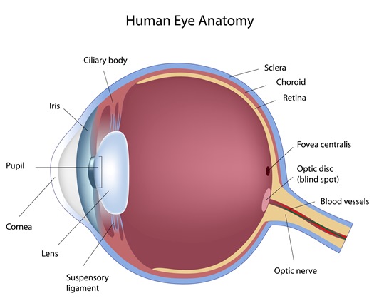
Cornea
The cornea is the dome shaped structure in the front part of the eye which covers and protects the rest of the internal structures of eye. The cornea is transparent, which allows you to see through it. It is comparable to the windshield of a vehicle, which is clear and allows you to see through it but also protects everything inside. In addition to protecting the internal structures of the eye, the cornea provides two-third of the focusing power of the eye. Some abnormal Corneal Conditions include pterygium, keratoconus, cornea dystrophy, Fuch’s corneal dystrophy, corneal ulcers and infections.
Crystalline Lens
The Crystalline Lens, also known simply as the lens, is a transparent structure in the eye found behind the iris. The iris is the colored structure in the eye found behind the cornea, and the central round opening in the iris is known as the pupil. The crystalline lens provides the fine tuned focusing power that allows you to see both far away and close up, which is also known as accommodation. Accommodation is achieved by changing the shape of the crystalline lens through the action of small muscles in the eye known as the ciliary muscles. If you compare the eye to a camera, the pupil is similar to the aperture of the camera and the crystalline lens is similar to the lens in a camera, which is used to focus images. A common example of crystalline lens pathology is a Cataract.
Vitreous Humor
The vitreous humor, or simply the vitreous, is a clear gel that fills the eye cavity from behind the lens to the retina, which lines the back of the eye. The vitreous is made up of mostly water but has a gelatinous consistency comparable to that of uncooked egg white. With time, the vitreous can shrink and pull away from the retina. This is known as a vitreous detachment, which can cause floaters in your vision. A vitreous detachment is usually a harmless age related change, but frequent, numerous floaters should be evaluated by your eye doctor.
Retina
The retina is a light sensitive tissue layer in the back of the eye. The retina is vascularized, which means it has blood vessels to supply vital nutrients for metabolic functions. The retina lines the inside of the back eye wall, also known as the sclera. The retina can be compared to a layer of film in a camera, which is important for the development of detailed images. Some examples of retinal pathology include Diabetic Retinopathy, Macular Degeneration, Retinal Tear and Retinal Detachment.
Optic Nerve
The optic nerve is the nerve which connects the eye to the brain. The optic nerve is also known as cranial nerve II. Images detected in the eye on the retina are transmitted from the eye to the brain by way of the optic nerve. The optic nerve can be compared to the cable which connects a digital camera to a computer. A common example of optic nerve pathology is Glaucoma.

